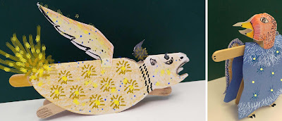
Workshop 1
Day 1, July 15th, 2019
Tashrīḥ-i badan-i insān or Mansur’s Anatomy manuscript
Housed at the Chester Beatty
Description of the manuscript: Manṣūr Ibn Ilyās was a late 14th century and early 15th-century Persian physician from Shiraz, known for his publication, Mansur’s Anatomy, of the first colored atlas of the human body. Mansur was born in the 14th century in the city of Shiraz, located in the providence of Fars in central Iran.
The manuscript consists of forty folios: an introduction, five chapters covering the osseous, nervous, muscular, venous, and arterial systems, and an appendix on the formation of the fetus and compound organs, such as the heart. This document led to a great deal of change in the way the Islamic world viewed human anatomy at the time. Mansur ibn Ilyas is also credited with one of the earliest anatomical sketches of a pregnant woman; while many believe his other illustrations to have been inspired by earlier Latin and Greek writings, the pregnant woman is considered an original work.
He discussed organs based on their hierarchical ordering of functionality-related groups according to their importance to the life of the body. In this manner, he discussed the anatomy of the vital and respiratory organs, and then the anatomy of the organs of nourishment, perception, and finally, reproduction. A concluding section on compound organs, such as the heart and brain, and on the formation of the fetus, was illustrated with a diagram showing a pregnant woman. Mansur's Anatomy is chiefly recognized for its inclusion of such colored anatomical illustrations, the first of its kind in medieval Iran and the Arabic regions.
History of anatomy in brief:
The final major
anatomist of ancient times was Galen, active in the 2nd century. The development of the study of anatomy
gradually built upon concepts that were understood during the time of Galen. Mundinus
in 14th C., performed the first human dissection recorded for
Western Europe. In 14th C. Mansur in Iran in the city of Shiraz published the first colored atlas of the human
body called, Mansur’s Anatomy. In Europe Vesalius was the first
to publish a treatise, De humani corporis fabrica, in 16th
C. that challenged Galen "drawing for drawing". These drawings were a
detailed series of explanations and vivid drawings of the anatomical parts of
human bodies. The study of anatomy flourished in the 17th and 18th
centuries. During the 19th century, anatomical research was extended and anatomical
research in the past hundred years has taken advantage of technological
developments and growing understanding of sciences to create a thorough
understanding of the body's organs and structures.
Curriculum:
Workshop Title:
Re-imagined Folios with Marginalia
Medieval Anatomy, Drawing, and Book-Page Layout
Medieval Anatomy, Drawing, and Book-Page Layout
Lesson Overview: Lesson Overview: Students learn about the
history of anatomy briefly and they explore anatomical concepts and images from
medieval Iran through pages of the Mansur’s Anatomy. Students also explore contemporary anatomy
through images; they compare the selected images from the two periods. Students
learn how to design their own manuscript pages with “marginalia”, how to draw
human bones (skeleton) and muscles. They write names of bones and muscles and
other relevant information next to their drawings. Students record their
thoughts about their exploration and discoveries as marginalia. After they
finished their pages, two students pair up to comment on each other’s work by
writing as marginalia. Note: Students can designate areas within the marginalia
for their peer’s comments.
Author: Pantea Karimi
Author: Pantea Karimi
Subject: Visual Arts, Science, Anatomy
Main Resource: Chester Beatty Library Archive
Workshop Time: Morning: 10:30 to 1:00 Afternoon: 1:45 to 4:15
Age Groups: Morning, 12-14 Afternoon: 15-17
Use of technology in the classroom: Students are given the opportunity to explore the human body through apps, and explore the internet for relevant information using provided tablets.
Art Vocabulary:
· Marginalia (or apostils) are marks made in the margins of a book. They maybe comments, critiques, drawings, or illuminations (decoration and miniature illustrations).
· Page layout is the part of graphic design that deals with the arrangement of visual elements on a page. It generally involves organizational principles of composition to achieve specific communication objectives.
Goals:
· Develop visual literacy and explore historical archives
· Develop a practicum based on the history of science through scientific manuscripts and
· Explore the intersection of art and science
· Use art as a tool to explore scientific content
· Effectively use the art and science resources offered by the Chester Beatty Library
Learning Objectives:
· Exercise and demonstrate the use of the elements of design
· Use materials, tools and processes from a variety of media (drawing, sculpting, and craft)
· Handle materials effectively
· Create original works of art in a specific medium using specific content
· Produce creative works that demonstrate innovation in concepts, formal language and/or materials
· Describe, analyze and interpret created artwork
· Demonstrate problem-solving skills by providing a step-by-step approach to specific issues in workshop projects
· Learn and explore science through the lens of art
Materials: Mixed-media paper preferably not smaller than 9x12 inches
pencil, eraser, scissors, ruler
Colored pencils, sharpies (black and assorted colors) or markers
Fasteners (for extra activity: skeleton puppets)
Copies of images for inspiration (refer to the end of this document)
Required time: 20 minutes: introduction- presentation by the teacher
10 minutes sketching and pondering ideas
2 hours of lab time (students develop their own works)
Project Steps:
Students receive 2 papers each, to create the manuscript
pages of anatomy.
Step 1: Paper 1, students sketch ideas for their page
layout and the composition of texts, skeleton, skull or other body parts. 5
minutes
Step 2: Paper 2: students divide the paper in half. They
draw a frame on each half and leave some margins for comments and recording
thoughts (marginalia) (preferably not smaller than 1 inch). 5 minutes
Step 3: Students draw images of human bones, skulls,
muscles, etc. inside the frames, they write names and other information in the
designated areas. 1hr:10minutes
Step 4: Students decorate around their frames if they
want to. 10 minutes.
Note: If they choose students can spend 10 extra minutes on their drawings
Note: If they choose students can spend 10 extra minutes on their drawings
Step 5: Students will record their thoughts and
discoveries about the human body as marginalia.
5 minutes
5 minutes
Step 6: Students pair up to comment on each other’s
work as marginalia after they are done with their manuscript pages. 5 minutes
See below images 1-15 from the Mansur's Anatomy manuscript, contemporary images of skeleton and muscles and Chester Beatty's students' works
See below images 1-15 from the Mansur's Anatomy manuscript, contemporary images of skeleton and muscles and Chester Beatty's students' works
Assessment:
Students will be assessed on:
· Use of creative page layout, design and marginalia
· Use of effective information and cohesiveness of their page layout
· Good drawing skills
· Good one-to-one discussion and use of marginalia for comments and recording thoughts
Images from the Mansur’s Anatomy and Vesalius's De humani corporis fabrica libri septem (Latin for "On the fabric of the human body")
image 1 Muscles and their descriptions from Mansur’s Anatomy manuscript
image 2 Image 1. Skeleton and description of bones from Mansur’s Anatomy manuscript

image 3, Vesalius Fabrica p194
image 4
image 5
image 6
image 7
image 8
image 9
image 10
image 11
image 12 (detail of image 11)
Photo by Chester Beatty, students at the anatomy workshop
image 13
image 14
image 15
Photo by Chester Beatty, students at the anatomy workshop


